Many of the immune cells that are important to our immune system develop in a small organ next to our heart: the thymus. However, with age, the thymus shrinks and the number of effective immune cells decreases. The Max Planck research groups from Freiburg and Würzburg have now identified the processes that control the growth and composition of thymus tissue throughout life. Furthermore, they have discovered new potential treatments to slow age-related shrinking of the thymus and fight autoimmune diseases.

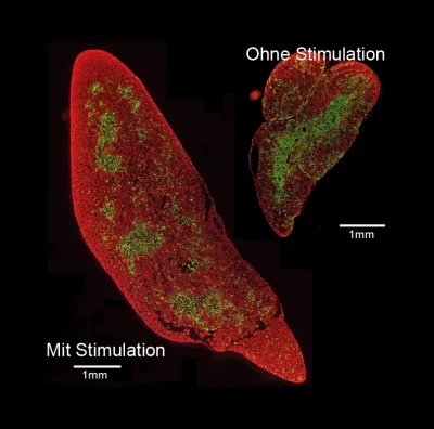
The smooth tissue structure of the stimulated thymus organ is not different from that of the unstimulated organ. As a typical sign of a well-functioning thymus, the marginal (red) and inner (green) regions are clearly separated from each other. Image credit: MPI of Immunobiology & Epigenetics, Boehm
The thymus is an important organ of the immune system. In the thymus, well-known T cells mature: As killer cells, they recognize and destroy virus-infected or malignant cells, and are called helper T-cells, they assist Helps the body to form antibodies. In the past decades, Thomas Boehm’s research group at the Max Planck Institute for Epigenetics and Immunobiology in Freiburg has identified genetic switches essential for T-cell maturation in the thymus. An essential component of this process is called thymic epithelial cells, which attract T-cell precursors and cause them to mature into fully functional T cells. During this development, T cells are instructed to distinguish diseased cells from healthy cells and foreign matter from the body’s own cells, thus allowing them to detect and eliminate Remove unwanted structures and prevent autoimmune diseases. Earlier studies at the Boehm laboratory had shown that two main types of thymic epithelium arise from pluripotent progenitor cells. However, it is not clear whether there is more than one ancestral form and it is not known how many subtypes the ancestors distinguish.
Molecular tree analysis identifies progenitor cells
Associate with Dominic Grün’s laboratory (formerly at the MPI in immunobiology and epigenetics in Freiburg, now the Max Planck Research Group at the University of Würzburg), an expert in single-cell RNA analysis, researchers have now succeeded in modeling describe the unexpected diversity of thymic epithelial cells at transcriptional levels. Algorithms developed in Grün’s lab to accurately characterize differences in the gene activity of individual cells help identify potential progenitor cells. In a second step, the researchers experimentally verified the predictions using a “barcoding” system developed in Thomas Boehm’s lab using CRISPR gene editing. The barcoding method allows the assignment of a molecular marker to progenitor cells, which are then carried by all cells emerging from the precursor. In this way, the researchers found a family tree of epithelial cells.
After a long period of method development marked by many failures, Anja Nusser from the Boehm laboratory and Sagar from the Grün laboratory succeeded together in developing a method of connecting information from the phylogenetic tree. species with the molecular features of individual cells. As a result, it was possible for the first time to study the development of the thymus epithelium at different ages in molecular detail. This type of analysis is of particular interest to immunologists because the thymus can change dramatically throughout life. Rapid organ growth and the production of large numbers of T cells are characteristic of the early stages of development. In contrast, there is a gradual loss of function of thymic epithelial cells with aging and, consequently, a decrease in T-cell production. These age-related changes are associated with decreased immune function.
The successive actions of progenitor cells determine the composition of the thymus
The researchers identified two biological precursor populations of the thymic epithelium in their analysis. An “early” progenitor population assumes a key role in thymogenesis during embryonic development. Whereas in juvenile organisms, a subsequent population of “postnatal” progenitors will significantly determine the continuation of thymus formation into adulthood. Interestingly, the chronological order of the prokaryotic population regulates the composition of the thymic epithelium.

Massive enlargement of the thymus organ is stimulated even in young mice, which is mostly preserved in old age. Image credit: MPI of Immunobiology & Epigenetics, Boehm
At baseline, mainly thymus epithelial cells are formed, contributing mainly to the production of T cells. At later time points, the major output is thymocytes. of the marrow. They ensure that no autoreactive T cells are released from the thymus into the body and thus make an important contribution to defense against autoimmunity.
New treatments to increase thymus function
The sophisticated combination of transgenic animal models from the Boehm laboratory with the Grün group’s state-of-the-art protozoan methods allowed the researchers to examine the effects of proliferating epithelial cells. thymus. It is particularly important to determine whether pre-stimulation of the thymus with a specialized growth factor results in faster stem cell consumption and thus premature thymic contraction. However, the data obtained by the researchers shows that this is not the case. “The thymus of old mice that were stimulated was still larger than that of young unstimulated mice. Furthermore, the fine-tissue structure of the stimulated thymus showed a typical structure of the cortical and medullary regions within the organ,” said Thomas Boehm, director of Max Planck. These results lay the foundation for the development of novel therapeutic approaches to correct age-related thymic shrinkage and to treat T-cell-dependent autoimmune diseases.
Source: MPG