Imagine that while you’re enjoying your morning bowl of Cheerios, a spider falls from the ceiling and rushes into the milk. Years later, you still can’t get close to a bowl of cereal without feeling disgusted.
Researchers have now directly observed what happens inside the brain learning that kind of emotional response. In a new study Published in January in Proceedings of the National Academy of Sciences, a research team at the University of Southern California was able to visualize memories forming in the brains of lab fish, visualizing them under a microscope as they expand in stunning fluorescent green. From previous studies, they had expected the brain to encode memory by slightly tweaking its neural structure. Instead, the researchers were surprised to discover a major overhaul in the connections.
What they saw reinforces the view that memory is a complex phenomenon involving a variety of encoding pathways. But it also further suggests that the type of memory may be crucial to how the brain chooses to encode it – a conclusion that may suggest why certain types of profoundly conditioned trauma responses are so persistent. so and it’s hard to learn.
“It’s possible what we’re looking at is the equivalent of a solid-state drive” in the brain, said co-author Scott Fraser, a quantitative biologist at USC. While the brain records some types of memories in a volatile, easily erased form, fear memories can be stored more strongly, which may help explain why years later some people can recall a memory as if it were coming to life, he said.
Memory is commonly studied in the cerebral cortex, which includes the top part of the mammalian brain, and in the hippocampus at the base. But it’s been less tested in deeper structures like the amygdala, the brain’s fear center. The amygdala is specifically responsible for associative memories, an important layer of emotional memories that link different things — like the spider in your cereal. Although this type of memory is very common, how it forms is not well understood, in part because it occurs in a relatively inaccessible region of the brain.
Fraser and his colleagues saw an opportunity to overcome that anatomical limitation and learn more about associative memory formation using zebrafish. Fish don’t have amygdala like mammals, but they do have a similar region called the pallium where associative memories form. While the developing mammal’s brain will grow by getting bigger and bigger – “inflating like a balloon” – the zebrafish brain almost turns inward, Fraser explains. popcorn kernels, so those deep centers are near the surface where we can visualize them. “What’s more, zebrafish larvae are transparent, so researchers can look directly into their brains.
Neuroscientists generally agree that the brain forms memories by regulating its synapses – the tiny holes where nerve cells meet. But most believe it mainly does so by regulating the strength of connections, or how intensely arousal one neuron is to the next, Fraser said.
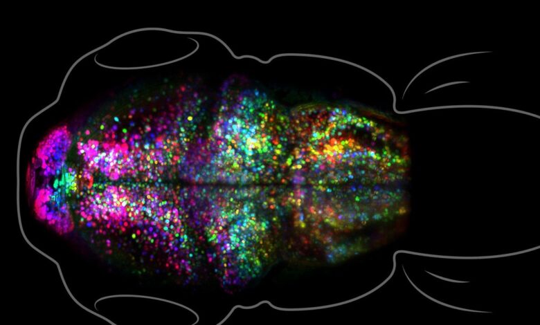
So to make that process clear, Fraser and his team genetically engineered zebrafish to create neurons with a fluorescent protein marker that binds to their synapse. . The marker protein, created in the lab of Don Arnold, a professor of biological sciences and bioengineering at USC, fluoresces under the dim laser light of a custom microscope. The challenge is “being able to eavesdrop on something while it’s happening,” but using as little light as possible to avoid scorching the creatures, Fraser said. The researchers were then able to see not only the location of each synapse, but also their strength – the brighter the light, the stronger the connection.